Microphytobenthos
analysis.
Three replicates of
1cm3 of sediment are collected from sediment cores. The replicates
are poured with 10ml of 2% glutardehyde. To avoid osmotic shock
formaldehyde solution was prepared on prefiltered seawater. In
laboratory, 0.5 to 1cm3 of two replicates were diluted in 10ml
of the 2% glutardehyde sollution and settled in sedimenting chambers
(10 ml volume). Each of the two replicates is analysed under the
inverted microscope. As minimum 500 cells is counted and identified
from each replicate. The third replicate was oxidised in 30% of
peroxide and mounted on Naphrax for qualitative analyses of diatoms
composition.
Phytoplankton & microplankton
Water column
Phytoplankton (microplankton)
is collected with the use of perplex glass bottle of 5dm3 volume
with closing device (Niskin Bottle or similar design). Samples
are collected from the depths delimited after the light measurement
(usually at surface, 50%, 10% and 1% of surface irradiance), in
the absence of light meter the samples are collected from 0, 2,
5,10 and 50m depth. Water sample onboard is placed into 250cm3
jar, fixed with Lugol solutiona and 3% formaldehyde. Samples are
analysed in the lab under the inverted microscope, after the sedimentation
in 1ml chambers. Phytoplankton is counted under the 200x magnitude
, and as minimum 1000 objects are identified and counted from each
sample.
Sea ice
Ice samples are collected
with the corer (drill) from the surface or by SCUBA diver from
below. The ice core of 5cm diameter is sliced into 5cm thick parts,
each one is placed in separate plastic zip bag. In the laboratory,
the ice core is placed in the 250ml filtered seawater, and left
in 4oC in cooler for slow thawing. After the melting is completed,
the sample is palced in the jar and fixed with Lugol solution and
3% of formaldehyde.
Sorting and counting procedures are the same as for the phytoplankton.
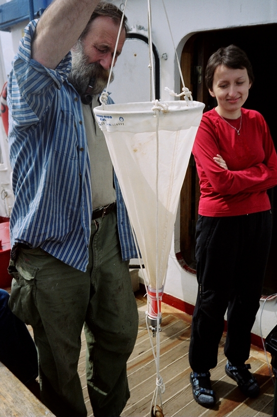
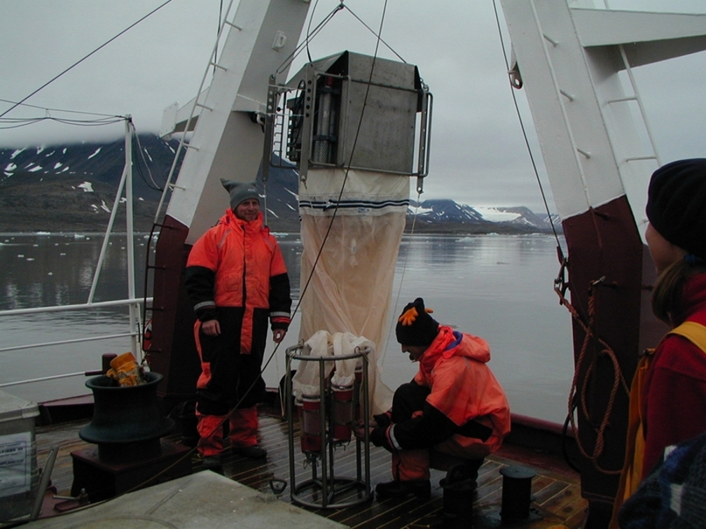
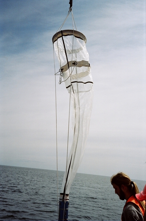
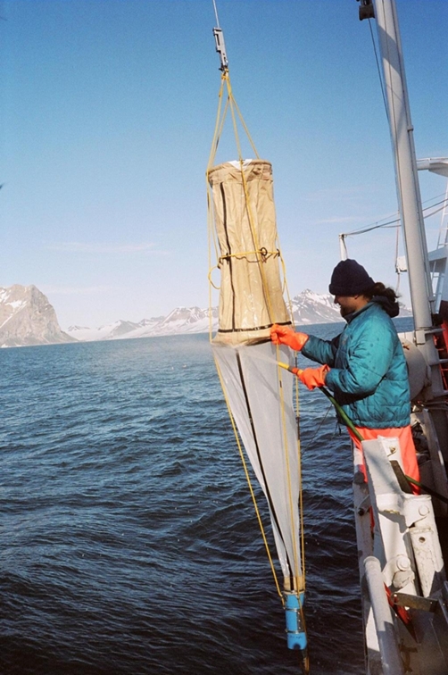
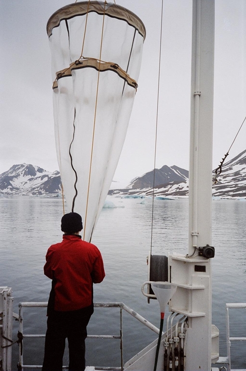
 Conical
net of 20 um mesh size for qualiatative
phytoplankton sampling Conical
net of 20 um mesh size for qualiatative
phytoplankton sampling |
 Cascade filtering
device with two different filters mounted;
upper filter with 10 um, lower with 0.4 um mesh size Cascade filtering
device with two different filters mounted;
upper filter with 10 um, lower with 0.4 um mesh size |
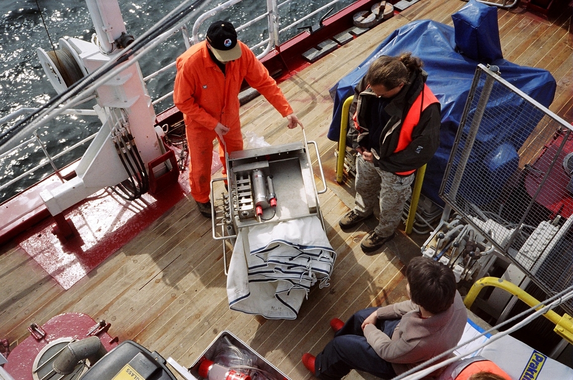
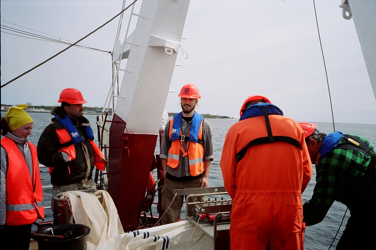
 Niskin
bottles, 5 and 15 l volume Niskin
bottles, 5 and 15 l volume |
|
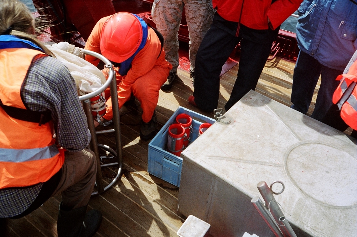
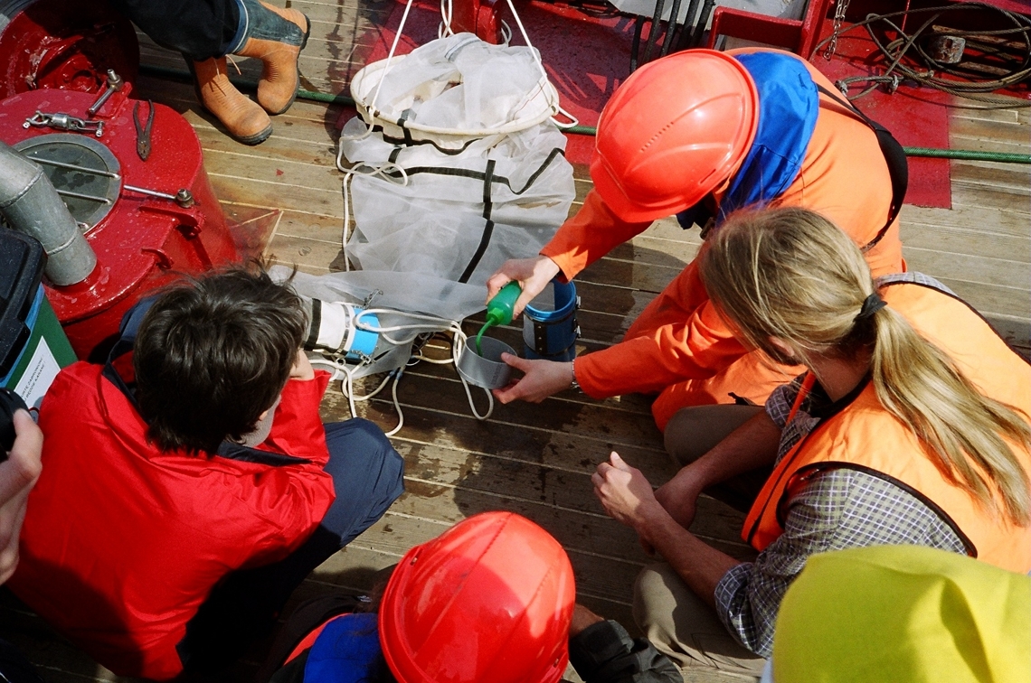
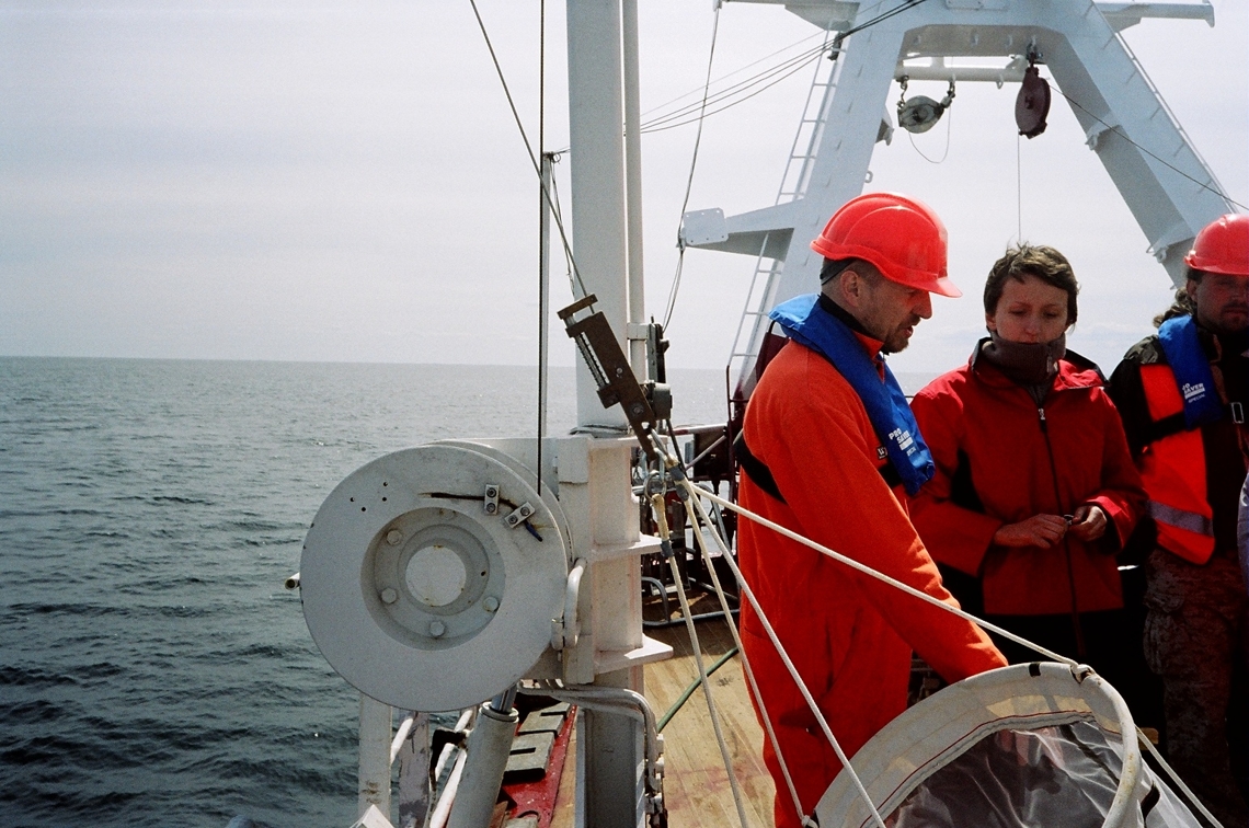
Mesozooplankton
Sampling
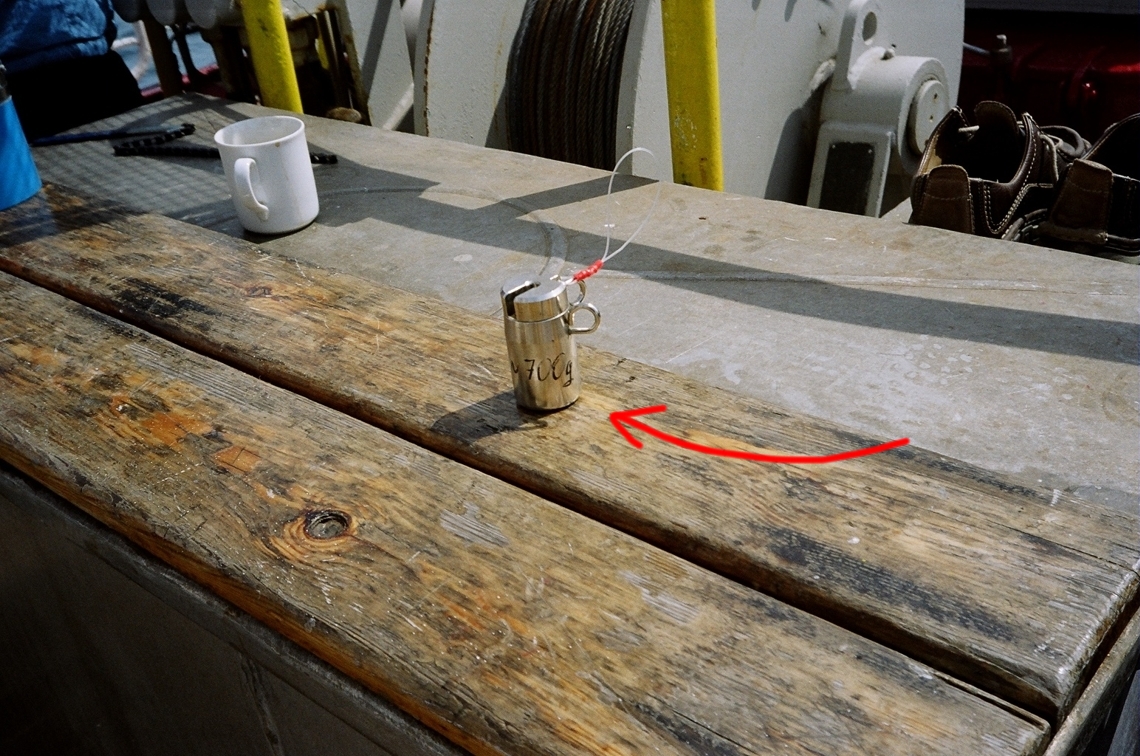
WP2 net with 57cm diameter,
180um gauze, closing device and flow meter is the standard net
used for vertical hauls. Routinely three water strata are sampled,
delimited after CTD measurements (usually: surface water - 0-20m,
intermediate layer - 20-50m, below picnokline - 50-200m). Vertical
sampling in layers thinner than 10m is not practical. Once retrieved,
the net is being washed externally with the hose with seawater
onboard, what allows flushing down the plankton attached to the
net column. Then the collector is removed and emptied into the
small plastic bucket. The collector's walls are rinsed gently to
remove all remaining plankton and the sample is poured into the
jar of 250ml volume. Excess water is being filtered out through
the same gauze. The sample is preserved with 37% formaldehyde buffered
with borax, to obtain 4% formalin solution in the plankton sample.
Second standard device is multinet sampler (Hydrobios-Kiel) with
0,25 m2 opening, equipped with 5 nets with 180um mesh size and
flow meters. Net is hauled vertically through the five discrete
water layers, established after the CTD profiling. The sample treatment
is the same as described above.
Sorting
In the laboratory, the
jar with zooplankton sample is placed under the ventilation funnel.
The sample is gently rinsed with tap water on the 180um gauze sieve
to remove the formalin. If needed sample is poured into the splitter
and divided into equal parts before analysis. Washed sample is
placed into100-200ml glass. All the conspicuous and large specimens
are removed first and identified. From the remaining plankton,
series of five subsamples are taken by 2 ml pipette and put into
the glass or plastic Petri dishes with an mm grid on the bottom.
Organisms from each sub- sample are identified, counted, and measured
under the stereomicroscope. At least 1000 small sized specimens
are identified from the sample. The rest of is looked through to
check for the large individuals, rare species and macroplankton.
Finally, the number of specimens counted is recalculated to the
original sample volume. After the identification, plankton is placed
back into the original jar.
Microfauna (Ciliates)
in sediments.
The sediment core is
obtained by inserting 36 x 297 mm plastic core into the sand, closing
with a rubber corks. The core is placed in the lab 30 minutes after
collection, and sliced in 2 cm sections to 20 cm sediment depth.
Subsamples for examination are chosen by inserting into each sediment
slice a 12 x 20 mm plastic mini-core, giving a subsample volume
of 2,26 ml. The subsamples are fixed in 10 ml 1% glutaraldehyde
solution (diluted with filtered water from the sampling location).
The samples are then stored in refregirator in 50 ml plastic containers
at 4°C.
Within two days from subsamples separation, the sediment slices are washed,
by shaking and then allowed to stand for approximately 5 s. A 5 ml sample of
water is removed with an Eppendorf pipette. The remaining sample is then supplemented
with adding 5 ml 1% glutaraldehyde solution, and after shaking another 5 ml
of supernatant is removed. The procedure is repeated 5 times, resulting in
25 ml of material from the washed sample. Assuming that ciliates were randomly
distributed in the solution immediately after shaking, 5 rinses removed 97%
of the ciliates present in the initial sample, because 0,55 x 100 = 3,1% of
the original sample remaining.
After shaking the obtained supernatant 10 ml is placed into plexi glass sedimentation
column for at least 6h. After this time settled ciliates are counted using
light microscopy at a magnification of 200x and 400x. Then cells are photographed,
measured and identificated to the lowest taxonomic level. Abundance is presented
in number of specimens per 2,26ml of sample. The biomass is calculated using
the conversion factors from the literature: 0,11 (ed. Edler 1979) and presented
in ug C/cm3.
For the examination of living material, two sediment cores are extracted and
sliced as described above, but not fixed. Sediment sections from cores were
slipped with minimal disturbance to a 50 ml plastic recipient and were immediately
carried to the laboratory. Extraction of the ciliates was made by the sea-water
ice method (Uhlig 1965). The cells were counted by removing them one by one
with a pipette under the dissection microscope and individual ciliates were
photographed using light microscopy at a magnification of 200x or 400x. Selected
ciliates were stained with protargol according to the method of Wilbert (1975.)
(Foissner et al.1999). A mixture of Bouin's fluid with saturated mercuric chloride
(1:1) was used as fixative. Photos and durable slides have been used to identify
species.
Literature:
Carey P.G. (1992) Marine Interstitial Ciliates. Chapman & Hall.
1-351.
Edler L.(ed.) (1979) Recommendations on methods for marine biological studies
in the Baltic Sea. Phytoplankton and chlorophyll. National Swedish Environment
Protection Board. 1-38.
Foissner W., Berger H., Schaumburg J. (1999) Identification and Ecology of
Limnetic Plankton Ciliates. Bavarian State Office for Water Management. 1-793.
Uhlig G. (1965) Untersuchungen zur Extraktion der vagilen Mikrofauna aus
marinen Sedimenten. Akademische Verlagsgesellschaft Geest &Portig
K.-G..151-157.
Meiofauna in
sediments
Meiofauna is collected
from the sediment cores, obtained from a perplex tubes of 3,6 cm
diameter (surface ~10 cm-2 is appropriate for all types of sediment),
inserted 15 cm deep into the seabed. From one site six (at least
three) replicates are taken.
Sediment is gently pushed out from the tube and cut into the slices. Te slices
are usually taken from particular layers: 0-1, 1-2, 2-3, 3-4, 4-5, 5-10 and
10-15 cm. Sometimes core is cut every 1 cm into even slices. Each sediment
slice (subsample) is placed into the separate jar, fixed with 4 % neutral formaldehyde
solution and stained with Bengal Rose, to obtain a pink/reddish color of the
sample.
For extraction of meiofauna from sediment are used two methods depending of
the amounts of the detritus or silt-clay in the sediment.
Decantation method - when the sediment is a sand with low amounts of detritus
or silt-clay.
In the laboratory, each slice is placed in the 1000 ml cylinder, filled with
tap water and shaken vigorously, to suspend the sediment grains. The water
is then filtered through 0.038 mm screen, and the procedure of shaking and
flotation is repeated 10 times. All meiofauna organisms are retained on the
screen, are gently washed into the Petri dish with measuring grid on the bottom
and counted under the low power stereo microscope.
Density gradient centrifugation method - the extraction from mud or detritus
is most efficiently using a density gradient in a centrifugation procedure
(Heip et al. 1985).
In this method liquid with a density larger than the density of meiofaunal
organisms can be used (Ludox, density of 1.15)
The method consist: placement of sediment sample into the Ludox solution, centrifuge
it with 1800 g for 10 min. The meiofauna organisms are retained in the 'gel',
once the sediment is on the end of the tube. Repeat centrifugation three times
more. This method does not work for heavy foraminifera, since their density
is often equal to the sediment grain.
In the lab, the meiofauna organisms are counted on the measuring grid, usually
1000 specimens per sample (sediment slice).
Basic handboks:
Heip C., Vincx M., Vranken G., 1985, The ecology of marine nematodes. Oceanographic
Marine Biology Annual Review 23: 399-489.
Vincx M., 1996, Meiofauna in marine and freshwater sediments. [In:] Methods
for the examination of organisms diversity in soils and sediments (ed G.S.
Hall). CAB INTERNATIONAL
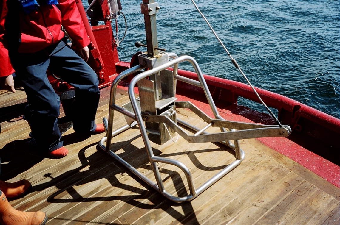
 Meiofauna
sampling - large syringes are inserted into box
corer sediment sample Meiofauna
sampling - large syringes are inserted into box
corer sediment sample |
|
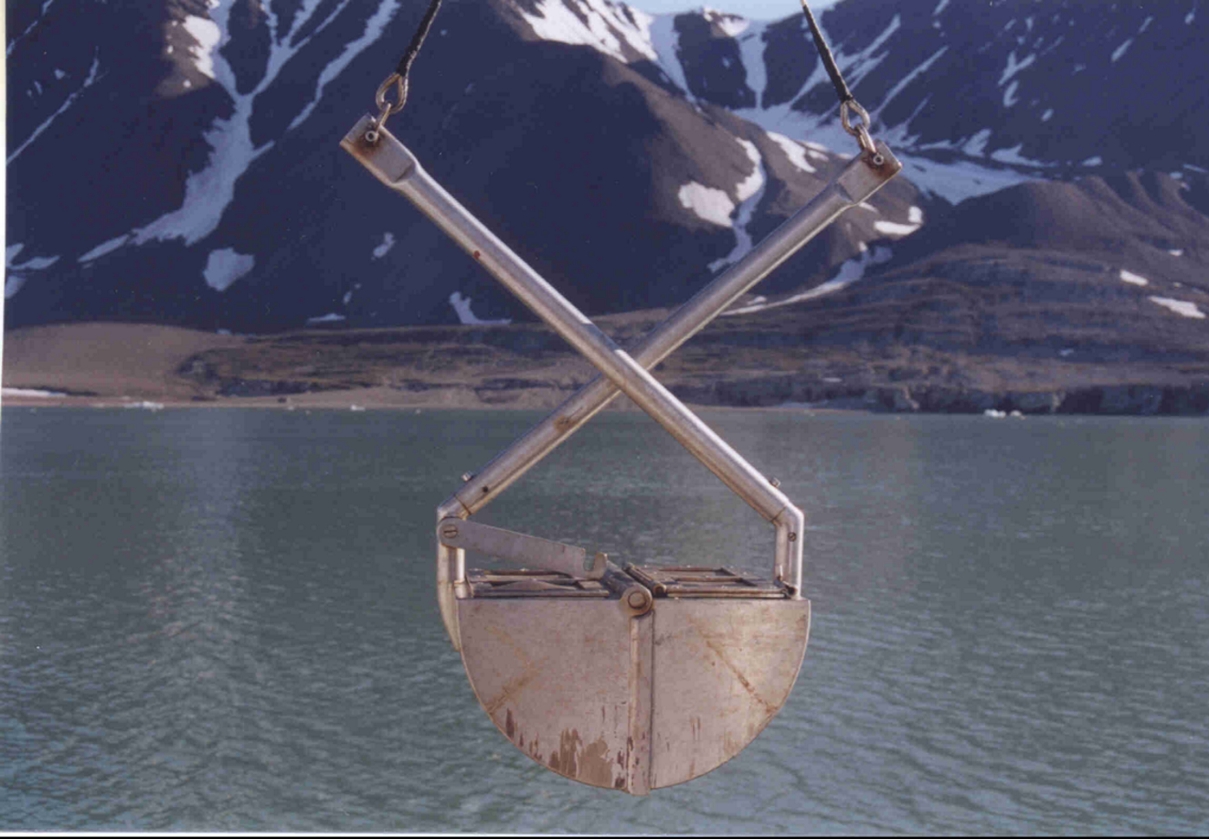
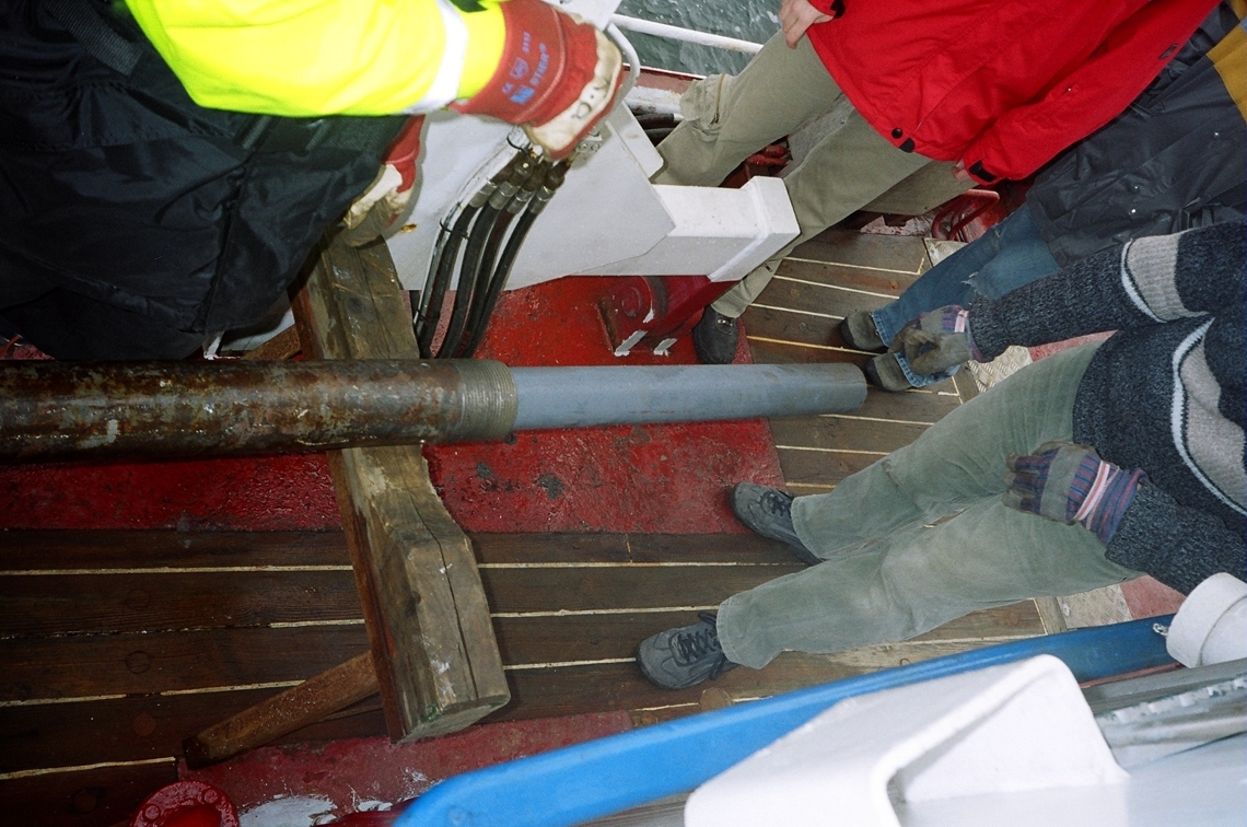
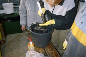
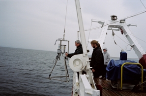
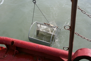
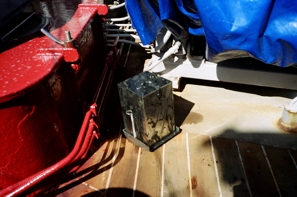
Macrozoobenthos
Sesile soft
bottom fauna, sublittoral - Van Venn grab, with flap
covers, 40kg weight, opening 30x30cm, triplicate samples from
one sampling point. After collecting the sample, first open the
grab onboard, using plastic spade remove gently upper 2cm of
sediment into the plastic 5dm3 bucket, wash gently with filtered
seawater and sieve on 0,5mm screen. Place the remaining sediment
into the washing tray, use gently filtered seawater from the
hoose and sieve organisms by flotation on 0,5mm screen. After
washing the sample, check large stones and gravel for the presence
of sessile organisms, remove clean rocks, and preserve organisms
in 4% formaldehyde solution in filtered seawater. In case of
substantial amount of sediment left use 10% formaldehyde solution.
In the laboratory sample is placed in the plastic container to
washout the formaldehyde, and is gently sorted again to remove
excess sediment. Organisms remaining on 0,5mm sieve are sorted
under the low power stereo microscope. From each sample all macrofauna
organisms are sorted, divided into separate jars containing main
taxonomic groups. All organisms are counted, for crustaceans
the body length is measured for each specimen (tip of rostrum
to telson), biomass (wet weight) is established after gently
blotting animal (or the whole number of specimens from given
taxon) on filter paper and weighted with 0,1mm accuracy.
Sessile hard
bottom fauna, sublittoral - SCUBA diving in the depth
of 2 to 30m, three randomly distributed metal frames (30x30cm)
on the selected type of habitat, within the same depth. All organisms
are collected (cut from the surface) from each frame into the
mesh bag of 0.5mm size. In case of motile organisms collection
form the hard bottom, the frame is equipped with gauze sac. The
ejector powered from the air bottle might be used for collection
of smaller, unattached animals hidden among rocks and stones.
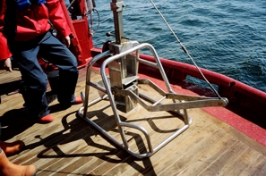
Motile epibenthos - light epibenthic sledge, equipped
with solid fabric apron, thick coarse net and 1mm mesh size gauze in
the cod end. The dredge is hauled along the previously recognized bottom
profile, within given depth interval, between 2m and 300m. Usual ratio
of a cable length needed to haul the dredge is 3 times the depth needed,
the hauling speed does not exceed 2 knots, and a haul duration range
from 10 to 30 minutes. The whole dredge is hauled out on the working
deck, and depending on the type of sample obtained (less or more muddy)
a subsample or the whole macrofauna is hand picked. The sample is a
qualitative one, giving only the rough proportion of the density of
organisms living on the seabed. Having number of dredge samples along
a transect, permits to get the good information on the macro faunal
species richness from the analysed area.
Carrion feeders - number of fast moving organisms
may avoid grabs and dredges or are too small to be collected into the
fishing gear like beam or otter trawl. Carrion feeding crustaceans
belong to the group of animals that are best collected with the use
of baited traps. The one used in our department is a cylindrical construction
of 1m per 30cm, covered with 1mm gauze, with two conical entrances
on both ends. The bait (100g piece of fish or bird’s meat) is placed
in nylon mesh bag inside the trap. Trap is lowered on the seabed with
an 2kg weight, and fastened to the buoy on the surface. Operational
depths are 2 to 200m, deeper on, the acoustic release system is practical.
The trap is exposed from 2hours up to 24 hours, and retrieved onboard.
The catch (may reach over 1kg of biomass) is removed from the opening
on the side of the net, washed in filtered seawater and placed in the
preservative. The position, amount of bait before and after the exposure,
depth, duration of exposure are noted.
Intertidal sedimentary bottom - quantitative sampling
is performed with the use of plastic tubes of 10cm diameter, inserted
at least 10cm into the sediment. Three to six replicates are collected
from any site, usually organized along the water marks - at Low Water,
Mean Water and High Water Mark. The coarser the sediment the deeper
the sample should be collected (in mud the upper 10cm, in coarse sand
the upper 40cm).
Intertidal hard bottom - quantitative sampling is
performed with the use of 20x20cm metal frame, placed on the selected
water mark during ebb tide. All organisms are cut, scraped from the
frame area and gently collected into the collector's jars. Three to
six replicates are recommended.
Basic handboks: Holme
AN, Mc Intyre AD. 1971. Methods for the study of marine benthos.
IBP Handbook no 16, Blackewll Sci. Publ. Oxford, 334pp
 Van
Venn grab Van
Venn grab |
|
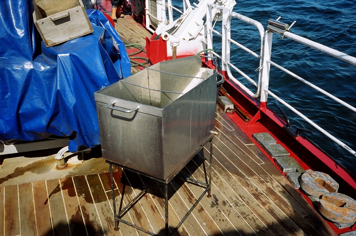
 Container
for washing sediment samples Container
for washing sediment samples |
 Small box
corer Small box
corer |
|
 Epibenthic
sledge Epibenthic
sledge |
 Camera
and video recording system ready for lowering Camera
and video recording system ready for lowering |
|
 Epibenthic
sledge Epibenthic
sledge |
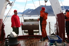
 Box
corer - removable box with newly collected
sediment Box
corer - removable box with newly collected
sediment |
|
|
Sedimentation
measurements
Suspensions
measurements
Water samples of 1.7 dm-3 are collected with the water sampler (Niskin bottle
type) from various depths, depending on the sampling design (usually 0m, 5m,
50m depth). Water subsamples of 100 to 500ml are filtered on combusted and
pre weighted Whatman Glass Microfibre filters GF/F, 0.7 um to get concentration
of Particulate Total Mater (PTM). Further procedures as described above with
sedimentation samples processing.
Gravity flows are measured one meter over the sea bottom with Sensordata currentmeter
Mini SD 6000 combined with Seapoint Turbidity Meter emitting light 880 nm and
scatterance angles 15-150 degrees. Empirical data of solids concentration from
Adventfjorden are then used to calibrate backscatter by plotting PTM (mg dm-3)
against FTU (Formazin Turbidity Units) recorded by Seapoit Turbidity Meter.
Received linear regression equation TPM = 0.9986 FTU+0.6899 (determination
coefficient R2= 0.999) was used to calculate concentration of PTM and gravity
flows.
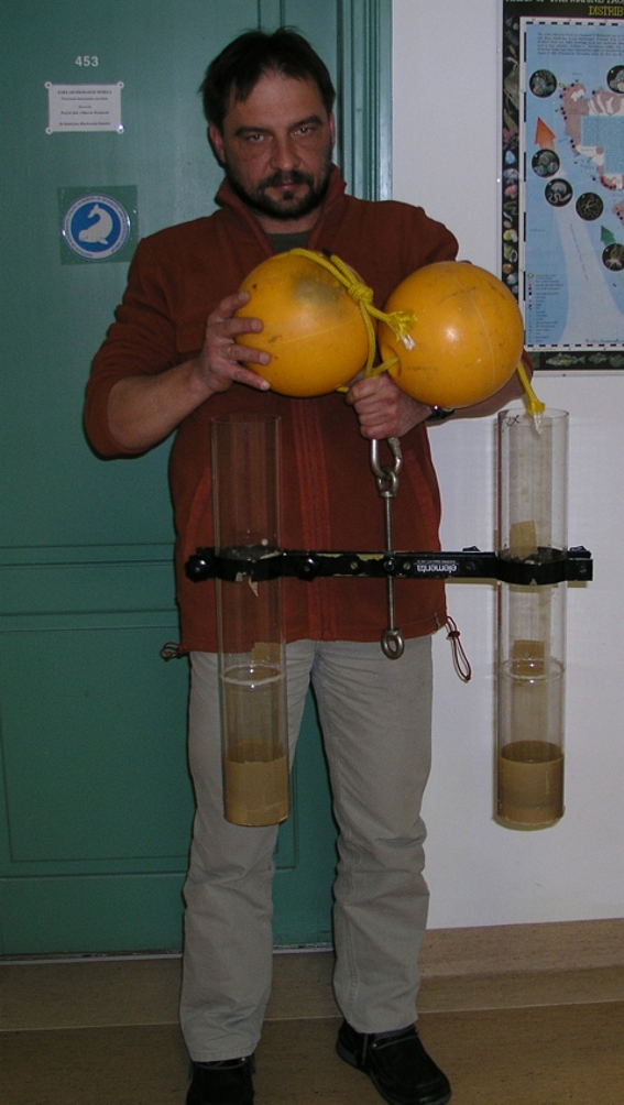
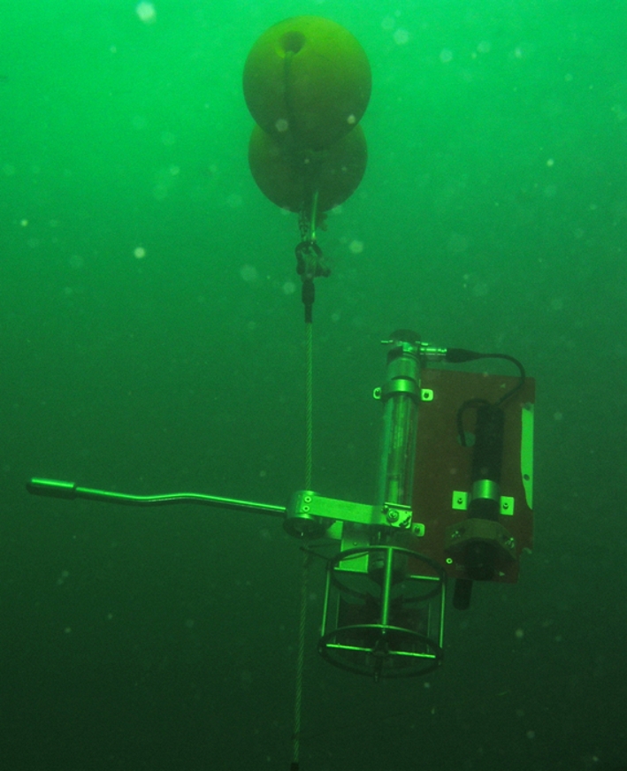
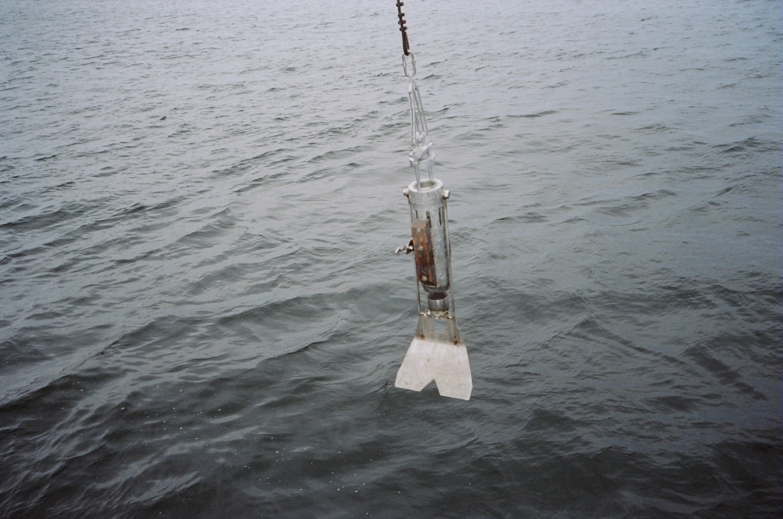
Sedimentation
measurements
The sediment traps are cylinders of perplex, 9.4 cm in diameter and 70 cm in
length or 10/100cm.. Equipped with the outlet 100 cm from the bottom. Traps
are deployed vertically in the water column, attached to the rope with buoy
in such way, that allow them to hang vertically (Zajączkowski 2002) Traps are
deposited from 12 to 48 hours, depending on the sedimentation intensity. Traps
are deployed below euphotic zone or below the pyknokline, 1m above the seabed
and usually in between in water column. Once retrieved, the samples is placed
onboard vertically, to avoid mixing the collected sediment, and the excess
water is removed through the outlet. Remaining 1 dm-3 of water with the accumulated
sediment is placed into the sampling jar. In case of delay between collection
and lab processing the sediment sample is fixed with 3% of formaldehyde.
In the lab, suspensions
subsamples are filtered in combusted and pre-weighted Whatman Glass
Microfibre filters GF/F 0,7um, usually 100 to 200ml are filtered.
After the filtration, the distilled water is used to wash out the
salt particles from the filter. Some filters are kept frozen at
-18 C for further analyses (maximum 12 days storage for chlorophyll
a), filters meant for the TSM (Total Sedimented Matter ) analyze
are dried in 60 C to stable weight, weighted, and next burned
in 450 C for 24 hours to combust the organic particles. After
the burning, the filters are weighted again, and the remaining
weight is considered as ISM (Inorganic Sedimented Matter). The
organic matter amount is calculated as a weight loss in combustion.
Additionally, subsamples of 50 to 100 ml of water are filtered
on combusted filters for particulate organic carbon (POC) and nitrogen
(PON) analyses. Before processing on Perkin Elmer 2400 CHN analyzer,
samples are stored 24 h in vapours of hydrochloric acid to remove
of calcium carbonate, and then ventilated 8 h (Hadges, Stern 1984).
Part of the original suspensions sample (usually 50 ml) is placed
in the glass cylinder for 24 hours for settlement of suspensions,
and after removing the excess water sediment sample is examined
under the microscope for particles indentification.
Literature:
Hadges, J.I., Stern J.H. 1984, Carbon and nitrogen determinations of carbonate
containing solids. Limnol. Oceanogr. Vol. 29: 657-663
Zajaczkowski M., 2002, On the use of sediment traps in sedimentation measurements
in glaciated fjords. Polish Polar Research. Vol. 23: 161-174.
Suspensions
measurements
Water samples of 1dm3
are collected with the water sampler (Niskin bottle type) from
various depths, depending on the sampling design (usually 0m, 5m,
50m depth). Water subsamples of 100 to 500ml are filtered on pre
weighted GFC filters of 045 um. Further procedures as described
above with sedimentation samples processing.
Sediment analyses.
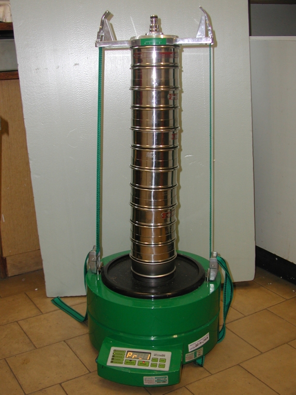
1. Granulometry table
with Wenworth scale of sediment size.
 The
sieves set with shaker The
sieves set with shaker
2. Organic matter
content
Organic matter content is measure in sediment samples of 1g dry mass, previously
dried in 65oC to stabilize weight. Sample is placed in the marked, pre-weighted
china jar in oven and burnt in 450 C for 24 hours. After the cooling of the
oven, the sample is weighted again in the jar, and the weight loss is considered
to be the organic matter loss in combustion. The organic matter content is
presented as the % of original dry sediment weight.
Organic Carbon Content in sediment - the samples of 1g dry sediment are analysed.
3. Porosity
Porosity represents the amount of water that may fill the given sediment unit
(mm3 of water per g of sediment)
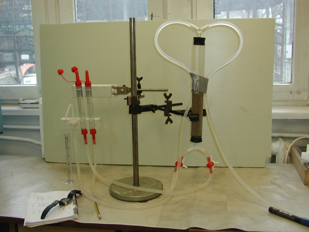
4. Permeability
Permeability presents the rate of water penetration through the sediment and
is expressed in mm3 per unit of length (cm) in time (sec).
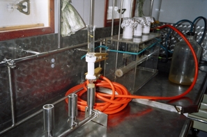
 The
equipment set for the permeability measurements The
equipment set for the permeability measurements
5. Chlorophyll
a content
Sand sample (surface 0,5cm of sand thickness, and 2cm in diameter) is placed
in the glass jar filled with 25 ml of distilled water, shaken vigorously for
15 minutes, and filtered through the GFC Whatman cellulose filter. Filter is
folded in half and frozen in -18oC, for no more than 14 days, within that time
the another extraction in acetone is performed, and the supernatant is analysed
in spectrophotometer 460 to 670 nm wavelength. Three sub-samples are taken
for spectroscopic meaurments.
Pore water sampling.
Pore water shall be
removed from distinct sediment layer, avoiding contamination of
sample from the water column. For the sediment depths up to 30cm,
different kinds of syringes are used, equipped with screen that
protect surface water from penetrating the sediment. For deeper
sediment (30-60cm) the peeper is used - double steel tube with
number of small holes along its length. Surface, fresh water outflows
is collected with the Bokuniewicz method - a barrel or tray with
attached plastic bag is digged into the sand and after 1-6hours
it collects the emiting freshwater.
Carbohydrate
concentration and composition (for sandy sediments).
I. Sampling:
1. Collect one sediment core (16cm length, 3.6cm diameter) from each following
depths: shore line, 1.5m and 7m
2. Slice each sediment core for 8 slices (samples): 0-2cm, 2-4cm, 4-6cm, 6-8cm,
8-10cm, 10-12cm, 12-14cm, 14-16cm
3. Freeze samples
II. Total Carbohydrate measurements:
1. Weight out 0,5g of each samples into test tubes. Record exact weight of
each sub-sample. For each sample should be weight out 3 sub-samples
2. Add to each sub-samples 1ml of dH2O
3. Prepare 3 blank: add to empty test tubes 1ml of dH2O
4. Add to each test tube 0.5ml of 5% phenol then quickly add 2.5ml of conc.
H2SO4, ensuring that the sub-samples are well mixed
5. Leave to cool for ~ 30min
6. Read in spectrophotometer at 485nm
III. Colloidal carbohydrate measurements:
1. Weight out 1.0g of samples into centrifuge tubes. Record exact weight of
each sub-sample. For each sample should be weight out 3 sub-samples
2. Prepare 3 blank: add to empty centrifuge tubes 1ml of dH2O
3. Add to each centrifuge tube 4ml of 100Mm EDTA and shake in a vortex mixer
4. Leave in a water bath at 25 C for 30min
5. Centrifuge for 15min at 4000g
6. Transfer 1ml of supernatant to test tubes (each sub-samples)
7. Add to each test tube 0.5ml of 5% phenol then quickly add 2.5ml of conc.
H2SO4, ensuring that the sub-samples are well mixed
8. Leave to cool for ~ 30min
9. Read in spectrophotometer at 485nm
IV. EPS carbohydrate measurements:
1. Transfer 1.5ml of supernatant from the colloidal fraction to centrifuge
tubes (each sub-samples and blanks)
2. Make up each of them to 70% ethanol (add 3.5ml industrial methylated spirit)
3. Leave overnight to precipitate at 4 oC (cover tubes to minimize loss)
4. Centrifuge at 4000g for 15min
5. Remove all supernatant and re-suspend EPS pellet in 1ml of dH2O
6. Transfer 1ml of each re-suspended EPS to test tubes (blanks too)
7. Add to each test tube 0.5ml of 5% phenol then quickly add 2.5ml of conc.
H2SO4, ensuring that the sub-samples are well mixed
8. Leave to cool for ~ 30min
9. Read in spectrophotometer at 485nm
V. Bounded carbohydrate measurement [with dH2O]:
1. Add to each of the pellet from the colloidal fraction 4ml of conc. ethanol
(blank too)
2. Shake in a vortex mixer
3. Centrifuge at 4000g for 15min
4. Remove all supernatant (if it is colored: even few times - until supernatant
will be colorless)
5. Add 4ml of dH2O and shake in a vortex mixer
6. Leave in water bath at 95 C for 1h, shake in a vortex mixer after 30min
of bath
7. Centrifuge at 4000g for 15min
8. Transfer 1ml of supernatant to test tubes (remove rest of supernatant)
9. Add to each test tube 0.5ml of 5% phenol then quickly add 2.5ml of conc.
H2SO4, ensuring that the sub-samples are well mixed
10. Leave to cool for ~ 30min
11. Read in spectrophotometer at 485nm
VI. Bounded carbohydrate measurement [with NaHCO3]:
1. Add to each pellet (bounded with dH2O) 4ml of 0.5M NaHCO3 and shake in a
vortex mixer
2. Leave in water bath at 95 C for 1h, shake in a vortex mixer after 30min
of bath
3. Centrifuge at 4000g for 15min
4. Transfer 1ml of supernatant to test tubes (remove rest of supernatant)
5. Add to each test tube 0.5ml of 5% phenol then quickly add 2.5ml of conc.
H2SO4, ensuring that the sub-samples are well mixed
6. Leave to cool for ~ 30min
7. Read in spectrophotometer at 485nm
VII. Standard Curve:
1. Dissolve 1g anhydrous glucose in100ml of dH2O (10mg/ml)
2. Take 1ml of 10mg/ml glucose solution and make up to 100ml with dH2O (100ug/ml)
3. Make 7 standard solutions:
a) 0ml of 100ug/ml glucose solution + 10ml of dH2O (0gu/ml)
b) 1ml of 100ug/ml glucose solution + 9ml of dH2O (10ug/ml)
c) 2ml of 100ug/ml glucose solution + 8ml of dH2O (20ug/ml)
d) 4ml of 100ug/ml glucose solution + 6ml of dH2O (40ug/ml)
e) 6ml of 100ug/ml glucose solution + 4ml of dH2O (60ug/ml)
f) 8ml of 100ug/ml glucose solution + 2ml of dH2O (80ug/ml)
g) 10ml of 100ug/ml glucose solution + 0ml of dH2O (100ug/ml)
4. Transfer 1ml of each solution to test tubes (3 samples for each solution)
5. Prepare 3 blank: add to empty test tubes 1ml of dH2O
6. Add to each test tube 0.5ml of 5% phenol then quickly add 2.5ml of conc.
H2SO4, ensuring that the samples are well mixed
7. Leave to cool for ~ 30min
8. Read in spectrophotometer at 485nm
|
 Department
of Marine Ecology, IO PAS, Sopot, POLAND
Department
of Marine Ecology, IO PAS, Sopot, POLAND